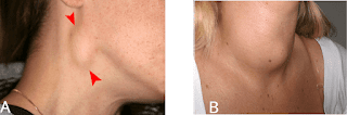
Eye tumor symptoms: What are they?

Breast Cancer: Watch Out For These Symptoms
By Charlotte Grainger of EspressoIt's time to talk about the C-word. Last year, there were more than two million new cases of breast cancer around the world, according to the World Cancer Research Fund. While anyone can be diagnosed with the disease, women need to be particularly on guard. In fact, the illness is now the most commonly occurring type of cancer in women.
One of the best ways to protect yourself is to perform self-examinations and keep an eye on warning signs. So, do you know the main symptoms of breast cancer and how to spot them? Let's take a look at 20 signs to watch out for.
© ShutterstockBreast Cancer Symptoms You Should Look Out For
Stay ahead of the trend in fashion and beyond with our free weekly Lifestyle Edit newsletterStay ahead of the trend in fashion and beyond with our free weekly Lifestyle Edit newsletter
Breast cancer is the most common cancer overall worldwide, and in the UK. According to the NHS, about one in eight women are diagnosed with breast cancer during their lifetime.
But a worrying number of women don't know the early signs of breast cancer. A 2019 study by cosmetics company Avon revealed that less than half (42 per cenr) of women surveyed were confident that they knew what changes to look for in their breasts.
Meanwhile, a quarter of women thought that a lump was the only sign of breast cancer. The findings were from a survey of 19,000 women. Only two per cent of those who participated could identify 10 common symptoms of breast cancer.
Although 73 per cent of participants said they regularly check their breasts, 60 per cent admitted they would be hesitant to seek medical advice out of embarrassment or fear.
In addition, the majority of women surveyed were unaware that their lifestyle choices could put them at greater risk.
Almost two thirds didn't know that regular exercise could help protect against cancer, while 63 per cent were unaware that alcohol is linked to a higher risk of the disease.
"Early detection is crucial to fighting breast cancer, yet our survey found that women don't know their risks or what signs to look for," said Sheri McCoy, chief executive of Avon.
Dr Paul Goss, chairman of the Avon Foundation scientific advisory board and Director of breast cancer research at Massachusetts General Hospital, added, "These figures show just how much work still has to be done in raising awareness of breast cancer, particularly its signs, risks and how to act on concerns about it."
So, what are the signs and symptoms that you should be looking for?
Women are advised to check their breasts each month - aside from a lump, other signs of breast cancer include a change in breast size or shape, a rash or skin sores, nipple discharge, skin indentation, constant pain or a change in skin texture.
Similarly, swelling around the armpit or collarbone could be an indicator, as can a growing vein or inverted nipple.
If you notice any symptoms of breast cancer, the NHS advises that you see your GP as soon as possible.
After examination, your GP will then refer you to a specialist breast cancer clinic if they feel your symptoms need further assessment.
You can find more information on how to check your breasts for cancer symptoms here.
Breast Lump Or Breast Changes: Early Evaluation Is Essential
© 2022 Mayo Foundation for Medical Education and Research (MRMER). All rights reserved. Breast and nipple changes can be a sign of breast cancer. Make an appointment with your health care provider if you notice anything unusual.Finding a breast lump or other change in a breast might cause worry about breast cancer.
That's understandable. But breast lumps are common. Most often they're noncancerous (benign), particularly in younger women. Still, it's important to have any breast lump evaluated by a health care provider, especially if it's new or if one breast feels different from the other breast.
How breast tissue feelsBreasts contain tissues of different textures, including fat, glands and connective tissue. Some breast-related symptoms, such as tenderness or lumpiness, change with the menstrual cycle. Lumps during this time might be caused by extra fluid in the breasts. Breast tissue also changes during pregnancy and menopause and while taking hormones.
When to consult a health care provider © 2022 Mayo Foundation for Medical Education and Research (MRMER). All rights reserved. A breast MRI requires lying face down on a padded scanning table. The breasts fit into a hollow depression in the table, which contains coils that detect magnetic signals. The table slides into the large opening of the MRI machine.Being familiar with how breasts usually feel makes it easier to detect when there's a change.
Reasons to consult a health care provider include:
Evaluation of a breast lump typically begins with a clinical breast exam. During this exam, a health care provider will likely:
© 2022 Mayo Foundation for Medical Education and Research (MRMER). All rights reserved. During fine-needle aspiration, a special needle is inserted into a breast lump and any fluid is removed (aspirated). Ultrasound -- a procedure that uses sound waves to create images of the breast on a monitor -- might be used to help place the needle.If the care provider finds a breast lump or other area of concern, you'll likely need testing.
Procedures to evaluate a breast lump Imaging testsTo further evaluate a breast lump, a care provider might recommend:
Newer tests for breast imaging are being developed and studied.
Breast biopsyThis involves having a tissue sample removed and examined under a microscope (biopsy). Ultrasound or mammography might help guide the needle, and a local anesthetic might be used. Breast biopsy options include:
After a biopsy, the tissue sample is sent to a lab for analysis. A health care provider explains the results.
Follow-up after breast lump evaluationIf the breast lump isn't cancerous, a health care provider will decide if there's a need for short-term monitoring with clinical breast exams or repeat breast imaging. You may be asked to return in 2 to 3 months to see if there have been changes in your breast. Consult your provider if you notice changes in the lump or develop new areas of concern.
A diagnosis in question might result in further consultation with a surgeon or other specialist. A diagnosis might be in question, for example, when a clinical breast exam and the mammogram show areas of suspicion, but the biopsy is benign.
A cancerous breast lump requires treatment. The tumor type and other factors will influence treatment options.
©2022 Mayo Foundation for Medical Education and Research (MRMER). All rights reserved.


Comments
Post a Comment